Blog
Dental hygiene tips for healthy teeth & gums
Types of Dental X-Rays
Most people know dental X-rays are a thing, but that’s about as far as it goes. Someone says an X-ray is needed, and it’s rarely questioned. A sensor gets placed. There’s a quick click. The chair comes back up. Everything just moves on. In the moment, it doesn’t feel important enough to stop and ask more.
Over time, things start to repeat themselves in a way people actually notice. Different visits involve different images. One X-ray reveals an issue that another missed. That’s usually when people begin to realise that different types of dental X-rays exist for a reason.
Why Dentists Don’t Rely On Just One X-Ray
Teeth are small, but what’s happening around them isn’t always simple. Cavities don’t always show on the surface. Bone loss doesn’t announce itself early. Infections can sit quietly for a long time before pain appears.
That’s why dentists use different views. One image can’t show everything. Each of the types of dental X-rays is designed to look at a specific area or problem. Some focus on teeth. Others focus on bone. Some capture fine details. Others give a broader picture.
Bitewing X-Rays And Why They’re So Common
Bitewing X-rays are usually the ones people recognise first. You bite down on a small tab. The image shows the top and bottom teeth together. It’s familiar, almost automatic, and usually over in seconds.
Dentists often use these X-rays to check between teeth, mostly because those spots are easy to miss. The spaces are narrow. Even good lighting doesn’t always help during a regular exam. A tooth can look completely fine on the surface while something is quietly developing below it.
Bitewings also give clues about bone levels. When dentists look at them over time, patterns start to show. Small shifts that wouldn’t stand out in a single visit become easier to spot.
That’s why bitewings end up being one of the most frequently used types of dental X-rays, even though they don’t always get much explanation.
Periapical X-Rays And What They Look For
Periapical X-rays are focused on one tooth at a time. They don’t just show the part you can see above the gums. The image goes all the way down to the root and into the bone around it, which is where a lot of problems tend to hide when something deeper is going on.
Dentists often take these images when pain has no obvious source. They’re used to look for infections, abscesses, root concerns, or shifts in the surrounding bone.
When someone hears that a tooth “looks fine on the surface,” but an issue still exists, a periapical X-ray is often the reason the problem gets identified. It plays a different role than bitewings, which is why it’s part of the broader types of dental X-rays conversation.
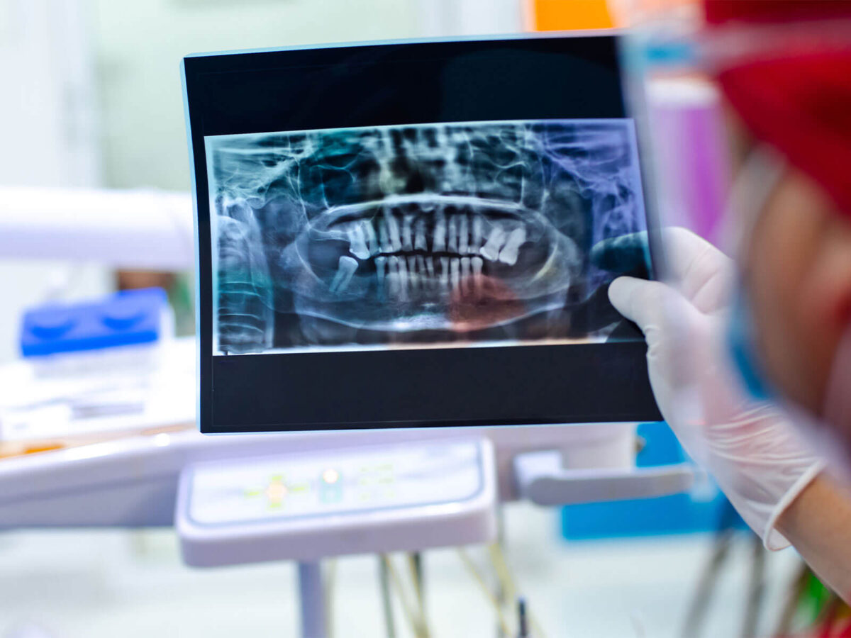
Panoramic X-Rays And The Bigger Picture
Panoramic X-rays work differently. They capture the entire mouth in one sweep. All the teeth are there, along with the jaws. Sinuses and joints are also included. It’s more about seeing everything together than zooming in on one spot.
These X-rays are not about small cavities. They are about the overall structure. Wisdom teeth. Jaw alignment. Impacted teeth. Bone abnormalities.
Because panoramic images show so much at once, they are used less frequently, but they are still an important part of the different types of dental X-rays dentists rely on for planning and diagnosis.
Occlusal X-Rays And Why They’re Used Less Often
Occlusal X-rays show how teeth line up within the jaw rather than focusing on individual teeth. The film is placed flat in the mouth, which already sets them apart from the X-rays most people are familiar with.
These images are usually used to find things that don’t show up easily otherwise. Impacted teeth. Extra teeth. Changes along the floor of the mouth. They aren’t part of routine care, so it’s common for patients to have never heard of them at all.
Even so, they serve a clear purpose. And that’s really the point behind all types of dental X-rays. Each one exists to answer a specific question.
Cephalometric X-Rays In Orthodontics
Cephalometric X-rays usually show up in orthodontics. Instead of zooming in on teeth alone, they show the side profile of the face. That wider view is what makes them useful in those cases. The jaw, the skull, and the overall structure all show up in one image.
These images help orthodontists understand growth and alignment issues. They’re not focused on cavities or restorations. They’re about positioning. Balance. How things fit together over time.
Most patients never need this type of X-ray. Even so, cephalometric images still fall under the umbrella of different types of dental X-rays, each one used for a very specific reason.
3d Dental Imaging And Modern Technology
Some dental offices use 3D imaging, often called cone beam CT scans. These images show teeth, bone, and nerves in three dimensions.
They are used for implant planning, complex extractions, and certain root canal cases. Because they involve more detailed imaging, they are not taken routinely.
This newer technology doesn’t replace other types of dental X-rays. It complements them when more detail is needed.
Why Not Every X-Ray Is Taken At Every Visit
Some patients worry when X-rays aren’t taken at a visit, or when the images look different from before. It can feel random if you’re not expecting it.
Most of the time, it isn’t random. Dentists look at what’s happening in the moment and then factor in what’s happened before. Past issues tend to guide what they pay attention to next. Frequent cavities usually mean more bitewings. Jaw pain often points to a panoramic image instead.
This is where understanding types of dental X-rays helps. It makes it clearer that imaging decisions aren’t automatic. They’re targeted.
How Often Dental X-Rays Are Needed
There isn’t a fixed rule for that. Some people need X-rays more often. Others don’t, and that difference is usually intentional. Age, oral health, and risk all play into that decision.
Dentists try to balance things. They look for enough detail to understand things, but they don’t take X-rays without a reason. Digital images use very low radiation now, but they’re still chosen thoughtfully. That same balance carries across all different types of dental X-rays.
Why One X-Ray Can Miss What Another Finds
Each X-ray captures a specific angle. Cavities between teeth may show on bitewings but not on periapicals. Jaw issues may show on panoramic images but not on close-up views. That’s why dentists don’t rely on a single image. Combining views creates a clearer picture.
What Dental X-Rays Do Not Show
X-rays don’t catch everything. Some problems just don’t show up well in an image. Early gum disease, small cracks, surface stains. Those often need to be seen directly or checked another way.
That’s why X-rays aren’t the entire process. They’re one tool among several. Understanding types of dental X-rays helps make sense of that, instead of expecting them to explain everything on their own.
Conclusion
So, the types of dental X-rays are not about taking more images than necessary. They are about choosing the right view for the right question.
Each image looks at something slightly different. When everything is looked at together, it helps dentists get a better sense of what’s happening beneath the surface. A lot of issues start there, well before they’re easy to spot or feel.
If questions come up around the different types of dental X-rays recommended for you, asking what each one shows can make things feel a bit clearer. Understanding why an image is taken often makes dental visits feel far more comfortable and informed.

